Heel spur causing heel pain

Posted on |

Heel spur causing heel pain

A heel spur is a bony growth that forms under the heel bone, and it can cause sharp or nagging pain and inflammation in the heel and its surrounding areas. Heel spurs are often caused by overuse, wrong shoes, improper foot mechanics, and certain medical conditions such as plantar fasciitis.

Heel pain from a heel spur is typically felt in the bottom of the heel and can be accompanied by:

  • Sharp pain when standing or walking
  • Pain that is worse in the morning or after prolonged sitting
  • swelling or redness in the heel
  • bony protrusion that can be felt or seen on the heel

Treatment for heel pain from a heel spur may include:

  • Rest and ice: Applying ice to the affected area can help reduce pain and inflammation.
  • Stretching and exercises: Stretching the calf muscles and Achilles tendon and doing exercises to strengthen the muscles in the foot and leg can help improve flexibility and reduce stress on the heel.
  • Orthotics: Customised shoe insoles can help correct any improper foot mechanics that may be contributing to the heel spur.
  • Medications: Non-steroidal anti-inflammatory drugs (NSAIDs) can help to reduce pain and inflammation.
  • Physiotherapy: A physiotherapist can treat this using Laser, Ultrasound and Radial Shockwave therapy and  exercises to improve strength, flexibility, and range of motion.
  • Surgery: In some cases, surgery may be recommended to remove the spur, but it is usually considered as a last resort.

 It is important to consult with a healthcare professional to determine the underlying cause of heel pain and the appropriate course of treatment. Additionally, some lifestyle modifications such as switching to shoes with a cushioned heel, avoiding high-impact activities, and incorporating low-impact exercises like swimming, cycling or yoga can also be beneficial in managing heel pain from heel spur.

You may contact us if you need help with your heel pain.


Foot and Heel

Plantar fasciitis is felt as a pain around the heel and arch of the foot. It can be felt as a discomfort or sharp pain in the heel on weight bearing especially after a rest period. As a person gets older, the fascia becomes less elastic. The heel pad becomes thinner and loses the capacity to absorb as much shock. There may be some swelling, small tears or bruises in the plantar fascia with the pounding force on the heel. Plantar fasciitis can also be a result of overuse in activities such as long-distance running, basketball, ballet dancing or dance aerobics. It settles down quickly if treated early and given enough rest, but may become worse and  chronic if initial symptoms are ignored.

To reduce the pain of plantar fasciitis, try these self-care tips:

  • Give adequate rest to your feet. Avoid prolonged standing or high impact activities like running that cause repeated loading on the foot. If you need to stand for long time, then shift your weight from one foot to the other or use a footrest under the affected foot to offload it for a while.
  • Don’t walk barefoot,especially on hard surfaces, as this puts extra stress on the plantar fascia. It is advisable to wear soft heeled footwear or footwear with scooped out heels to avoid pressure on the heel.
  • Wear supportive shoes.Choose shoes with a low to moderate heel, supportive arches and good shock absorbency.
  • Avoid high heels especially when you need to walk long distances or stand for long periods of time. High heel shoes exert additional pressure on the inflamed fascia and lead to more heel pain.
  • Do not wear worn-out shoes.Replace old, tattered, non-supportive shoes. This is very important if you walk or run in these shoes. A good way to tell that your shoes need replacing is to look for thinned (worn) out areas on the sole of the shoe.
  • Apply ice: This can be done on the painful area three or four times a day, especially at the end of the day. Icing helps to reduce pain and inflammation. Icing can also be done with a frozen bottle of water rolled under the foot while sitting.
  • Massage: Self massage can be done by rolling a tennis ball under your foot while sitting. As mentioned above, a frozen water bottle can also be used.
  • Change your sport.Try a low-impact sport such as swimming or bicycling instead of walking or jogging while the plantar fascia is inflamed/painful.
  • Maintain a healthy weight. If you are overweight, then try to lose some weight. Extra weight can put extra stress on your plantar fascia.
  • Exercise before getting out of bed in the morning or after prolonged sitting(sit to stand): Plantar fasciitis pain is usually at its worst in these two situations. A good way to combat this is to perform circular movements at the ankle (clockwise and anticlockwise) and a few seated calf stretches before weight bearing on the feet.
  • Do your stretches.Simple home exercises can be done for plantar fasciitis. Perform this stretch when waking up, mid-day, and before bed. It is also very important to perform these stretches in the warm up and cool down phase of your exercise routine, even after you recover from plantar fasciitis pain. This will help to prevent any recurrences. 

Heel Spur Causing Heel Pain

Description/Introduction:

A heel spur is a bony growth that forms under the heel bone, and it can cause sharp or nagging pain and inflammation in the heel and its surrounding areas. Heel spurs are often caused by overuse, wrong shoes, improper foot mechanics, and certain medical conditions such as plantar fasciitis.

Heel pain from a heel spur is typically felt in the bottom of the heel and can be accompanied by:

  • Sharp pain when standing or walking
  • Pain that is worse in the morning or after prolonged sitting
  • swelling or redness in the heel
  • bony protrusion that can be felt or seen on the heel

Treatment for heel pain from a heel spur may include:

  • Rest and ice: Applying ice to the affected area can help reduce pain and inflammation.
  • Stretching and exercises: Stretching the calf muscles and Achilles tendon and doing exercises to strengthen the muscles in the foot and leg can help improve flexibility and reduce stress on the heel.
  • Orthotics: Customised shoe insoles can help correct any improper foot mechanics that may be contributing to the heel spur.
  • Medications: Non-steroidal anti-inflammatory drugs (NSAIDs) can help to reduce pain and inflammation.
  • Physiotherapy: A physiotherapist can treat this using Laser, Ultrasound and Radial Shockwave therapy and exercises to improve strength, flexibility, and range of motion.
  • Surgery: In some cases, surgery may be recommended to remove the spur, but it is usually considered as a last resort.

 It is important to consult with a healthcare professional to determine the underlying cause of heel pain and the appropriate course of treatment. Additionally, some lifestyle modifications such as switching to shoes with a cushioned heel, avoiding high-impact activities, and incorporating low-impact exercises like swimming, cycling or yoga can also be beneficial in managing heel pain from heel spur.

You may contact us if you need help with your heel pain.

TREATMENT for PAIN IN NECK IN SINGAPORE

Description/Introduction

Bending on your computer or phone? Is your back is slumped right now? Are your shoulders rounded? Is your chin going too far forward? These are common postures to be seen now a days with the world of technology and gadgets, we prefer to slump with gravity as it’s an effort to sit otherwise.

Hence lots of us suffer from neck aches and neck pains resulting from the posture of tilting you’re the head down to look at the screens and devices. We tend to spend many hours a day sitting in these kind of postures hunching over the gadgets, whether it’s for work or leisure .

Causes of Neck Pain:

When you tilt your head forward or look down, this increases the weight of the neck and pressure on the neck muscles, soft tissues, and cervical vertebrae. 

Neck muscles have to work to hold the head up, and the more you look down, work gets hard for muscles work to keep your head up. Over time, the repetitive strain results in soreness, stiffness, and even injury to the neck muscles and cervical spine.

Clinical Signs and Symptoms of Neck Pain:

  • Ache or pain in the neck, shoulders, and upper back.
  • Sharp, stabbing pain in the same areas.
  • Stiffness. 
  • Increased neck and shoulder pain when looking down. 
  • Tingling, numbness, or weakness in the hands —spinal nerve pain
  • Temporomandibular joint (TMJ) problems
  • Cervical spondylosis

Tips by Physio Asia, an experienced physiotherapy centre in Singapore

  • Keep your screens at eye level. 
  • Invest in good supportive chair with a great headrest and lumbar support.
  • Strengthen and stretch your muscles. Learn correct exercise from the physiotherapist
  • Take frequent breaks to get up, walk around, and stretch for a minute every 15-30 minutes to help ease tension in the neck and body.
  • Correct your posture. Stand straight with your head centred over your neck and keep a neutral spine, so your ear is in line with your shoulders. 
  • Apply ice. You can apply ice for the first 48 to 72 hours to relieve pain in a stiff and sore neck. Then switch to warmth with a heating pad, hot compress, or warm bath to heal and stretch your muscles and tendons.
  • Visit our physiotherapist to help you relieve the pain

Try and follow preventative methods to prevent the aches and pains associated with posture. If your symptoms do not improve, you should seek help from a qualified health professional for neck pain treatment in Singapore. Call us for an appointment or send your queries to: info@physioasia.com

Real Time Ultrasound for Pelvic Floor Muscles or Diastasis Rectus

Real Time Ultrasound Imagery (RTUS) uses sound waves to provide a visual image of muscle, tissue, and ligaments in our bodies. RTUS is a not a treatment tool but is a diagnostic and learning modality, that allows the physiotherapist and visual learner a picture and active image of parts of their body and their functioning.

RTUS is a wonderful tool to view the Diastasis Rectus (DR) abdominal muscle separation that may occur because of pregnancy, trauma, birth defect and or obesity. Measurements can be taken with RTUS and an image stored to view later, to evaluate the success of the physiotherapy program provides great motivation and feedback for both the client and physiotherapist.

RTUS is painless, noninvasive and a fabulous diagnostics tool to allow physiotherapist trained in this area, a platform from which to create an individualized educational exercise or training program for our clients.

In the post-partum period, when mums are very concerned about returning back to exercise, and what kinds of eerie they should be doing – the use of RTUS allows for a clear visual image of their DR separation and from there a return to core fitness and a fun exercise program can be tailored for the client.

RTUS is also a fantastic tool to allow women to view their Pelvic Floor Muscles (PFMs) to see whether the PFMs are working affectively again and to see if any dysfunctions such as changes in muscle tone and recruitment patterns are noted.

RTUS can be used safely while pregnant, as well as after you have delivered your baby. Please come in with a semi full bladder, as this allows a clear view of the PFMs, no vaginal probe will be used. A RTUS sound head will be placed on your skin in the lower pelvic area to view your bladder and PFMs. This is completely noninvasive and painless. Everyone usually really enjoys seeing their body muscles and how they function from the outside in!

CERVICAL SPONDYLITIS OR SPONDYLOSIS

Cervical spondylosis, also known as cervical osteoarthritis or cervical spondylarthrosis is a natural age-related wear and tear (degeneration) process of the neck. It can involve the bonesdiscs, and joints of the neck.

With age or with over use in wrong postures ( at an early age too) as the cartilage wears out, the discs loose their original volume and can become dried and develop cracks and eventually become stiffer.  If left unchecked they can further develop into disc herniation, commonly known as slip disc.

Cervical spondylosis can also lead to ligament thickening and formation of bone spurs (osteophytes) in the areas where the bones rub against each other due to the lack of top layer of protective cartilage. All of these changes can lead to narrowing of the spinal column or the openings where spinal nerves exit, leading to a condition known as spinal stenosis.

Symptoms of cervical spondylosis:

Cervical spondylosis usually occurs in middle-aged and elderly people. One or more of these symptoms may be seen:

  • Neck pain
  • Stiffness in the neck
  • Pain or tingling numbness may radiate to the arm, forearm and/or hands and fingers.
  • Muscle weakness in the upper limbs.
  • Headaches, originating from the back of the head.

However, these symptoms can be easily managed by maintaining a good posture and maintaining flexibility and strength of the neck muscles.

But if the symptoms get worse it is always better to address them with a physiotherapy sessionPhysiotherapy can help to manage neck pain and stiffness and other symptoms caused by cervical spondylosis. Physiotherapy treatments can help patients achieve improvements such as reduction of pain, increased neck movements, increased neck strength, improved posture and therefore helps to return to activities of daily living and eventually potential return to sports.

Our physiotherapists will assess your neck and treat the presenting symptom with a combination of therapies, some of which are mentioned below:

  • Manual therapy such as mobilisations
  • Tissue releases
  • Laser
  • Ultrasounds
  • Short wave diathermy
  • Different current forms
  • Postural and ergonomic education are also a vital part of the treatment.

Home treatment includes exercises for stretching the tight muscles, strengthening exercises for weak muscles and use of cold packs to ease the inflammation.

We are also well versed with specialized treatment techniques such as:

  • Dry needling
  • Decompression therapy
  • Radial shock wave
  • SCORES – Spinal Core Stability Program for muscle strengthening and diaphragmatic breathing control, to strengthen the deep stabilising muscles of the spine.
  • Achievo pressure feedback system – This provides an audio-visual indication of whether you are working the correct postural muscles in the right manner when performing specific core exercises.

These specialized treatment techniques give an added advantage to the patient as they help to fasten healing and thus speed up the recovery time.

Plantar Fasciitis or Pain in Sole of Foot – Tips from our Physiotherapist

Plantar fasciitis is felt as a pain around the heel and arch of the foot. It can be felt as a discomfort or sharp pain in the heelon weight bearing especially after a rest period. As a person gets older, the fascia becomes less elastic. The heel pad becomes thinner and loses the capacity to absorb as much shock. There may be some swelling, small tears or bruises in the plantar fascia with the pounding force on the heel. Plantar fasciitis can also be a result of overuse in activities such as long-distance running, basketball, ballet dancing or dance aerobics. It settles down quickly if treated early and given enough rest, but may become worse and chronic if initial symptoms are ignored.

https://physioasia.com/foot-and-heel/

To reduce the pain of plantar fasciitis, try these self-care tips:

  • Give adequate rest to your feet.  Avoid prolonged standing or high impact activities like running that cause repeated loading on the foot. If you need to stand for long time,then shift your weight from one foot to the other or use a footrest under the affected foot to offload it for a while.
  • Don’t walk barefoot, especially on hard surfaces, as this puts extra stress on the plantar fascia. It is advisable to wear soft heeled footwear or footwear with scooped out heels to avoid pressure on the heel.
  • Wear supportive shoes. Choose shoes with a low to moderate heel, supportive arches and good shock absorbency.
  • Avoid high heels especially when you need to walk long distances or stand for long periods of time. High heel shoes exert additional pressure on the inflamed fascia and lead to more heel pain.
  • Do not wear worn-out shoes. Replace old, tattered, non-supportive shoes. This is very important if you walk or run in these shoes. A good way to tell that your shoes need replacing is to look for thinned (worn) out areas on the sole of the shoe.

  • Apply ice: This can be done on the painful area three or four times a day, especially at the end of the day. Icing helps to reduce pain and inflammation. Icing can also be done with a frozen bottle of water rolled under the foot while sitting.
  • M assage: Self massage can be done by rolling a tennis ball under your foot while sitting. As mentioned above, a frozen water bottle can also be used.
  • Change your sport. Try a low-impact sport such as swimming or bicycling instead of walking or jogging while the plantar fascia is inflamed/painful.
  • Maintain a healthy weight. If you are overweight, then try to lose some weight.  Extra weight can put extra stress on your plantar fascia.
  • Exercise before getting out of bed in the morning or after prolonged sitting (sit to stand): Plantar fasciitis pain is usually at its worst in these two situations. A good way to combat this is to perform circular movements at the ankle (clockwise and anticlockwise) and a few seated calf stretches before weight bearing on the feet.
  • Do your stretches. Simple home exercises can be done for plantar fasciitis. Perform this stretch when waking up, mid-day, and before bed. It is also very important to perform these stretches in the warm up and cool down phase of your exercise routine, even after you recover from plantar fasciitis pain. This will help to prevent any recurrences. 

1. Standing calf (gastrocnemius) muscle stretch:

  • Stand on the edge of a step (stairs) or stable stool
  • Lower one heel over the edge of the step
  • You should feel the stretch on the calf muscle.
  • Hold stretch for between 12-15 seconds, 3-5 repetitions each side.

2. Seated calf stretch:

  • Sit with your legs extended out in front of you and then bring your foot in towards you from the ankle at about 90 degrees.
  • You can get an additional stretch by using a towel or yoga belt and placing it around the ball of the foot.
  • You can either stretch one calf at a time or both together.

3. Soleus Stretch:

  • To stretch place the leg to be stretched behind and lean against a wall, keeping the heel down
  • A stretch should be felt lower down near the ankle at the back of the leg
  • Hold for 12-15 seconds, 3-5 repetitions each side.

4 . Stretching the deep foot flexors

  • In sitting position, gently hold foot with one hand
  • With the opposite hand pull all five toes up towards the body
  • Hold for 12-15 seconds, 3-5 repetitions each side.

5. Towel lifts

  • This is to strengthen the deeper muscles of the foot (intrinsic muscles)
  • Place a hand towel on the floor. The towel should be completely flat
  • Stand with your foot over the towel and use your toes and the bottom of your foot to scrunch up the towel
  • Next use your toes and feet to flatten the towel
  • Repeat 10 times, 2-3 times daily 

These are some of the ways in which plantar fasciitis can be managed. You can consult one of our SingaporePhysiotherapistif you continue to suffer from heel pain or if you need assistance with the exercises to take care of your heel pain. Call us for an appointment or send your queries to: info@physioasia.com

Tips to manage tennis elbow

Tennis elbow or pain on outer side elbow of the elbow is the most common overuse injury in the elbow. Tennis elbow is also known as lateral epicondylitisHowever the condition is not restricted to tennis players. It can occur in anyone who does repeated movements leading to overuse of extensor tendon, such as people who spend considerable amount of time typing and using the mouse without proper positioning of the wrist.

Self management includes stretching of the forearm extensor muscles within the pain free range. Some of the simple stretches that can be done for tennis elbow are given here:

Forearm stretch using your other hand:

  • Make a closed fist with the thumb placed inside the fist.
  • Extend the wrist and place the other hand on the fist to apply overpressure.
  • Feel the stretch on the forearm muscles.
  • Hold for 5-10 seconds (as per tolerance), repeat 10 times in the day.

  • Forearm stretch:
  • Place both hands together as shown in the picture.
  • Try to bring the outer part of the hand together as much as possible, all the way from the wrist to the fingers.
  • Hold for 5-10 seconds (as per tolerance), repeat 10 times in the day.

  • Forearm stretch against the wall:
  • Place the back of your hand against the wall, such that it is at right angles to the wall.
  • Feel the stretch on the forearm muscles.
  • Hold for 5-10 seconds (as per tolerance), repeat 10 times in the day.

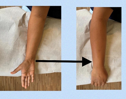
Forearm rotation:

  • Place your forearm at right angle to the arm.
  • Start with your palm facing upwards.

Turn the forearm such that the palm is facing downwards

Triceps stretch:

  • This stretch is done if you can feel the pain from the elbow going up into the back of the arm (into the triceps).
  • Keeping your elbow bent try to touch your upper back with the hand.
  • Use the other hand to apply additional stretch to the triceps.
  • Hold for 5-10 seconds (as per tolerance), repeat 10 times in the day.

Some other tips that can help to ease the pain and discomfort from tennis elbow are given below:

A good posture is vital to maintain the joints in neutral so that there is no excessive pressure on the joints. If you spend a considerable amount of time working on a computer, then use a mouse pad or ergonomic mouse so that your wrist is in neutral position (not bent upwards) while using a mouse.

Elbow brace can be used to alleviate pain by providing support to the muscles to stabilize the area.

Cold packs to settle the inflammation. Ice packs can help to minimize swelling around the elbow and reduce muscle spasm and pain.

If you participate in a racquet sport, check your racquet for a proper fit. Stiffer racquets and looser-strung racquets often can reduce the stress on the forearm, which means that the forearm muscles do not have to work as hard. Check the width of the racquet handle. If it is too thin it will lead to overgripping, leading to overuse of the extensor muscles.

Adequate warm up before and after exercise by gently stretching your elbow and arm muscles.

Our physiotherapists can help you to manage your tennis elbow and assist you with exercises to take care of any pain or complications arising from your tennis elbow. Call us for an appointment or send your queries to: info@physioasia.com

Hand pain – Occupational therapy for hand

wrist pain

Due to change in nature of work in recent times, most people working from home with different postures, uncomfortable work desks, long work hours, excessive use of phones and computers, hand, elbow pains are quite common occurrences.

Hand therapy is a specialized healthcare service provided mainly by occupational therapists which focuses on the rehabilitation of the upper limb (elbow, forearm, wrist, hand, fingers and thumb).

The hand is one of the most important parts of our bodies, responsible for completing most of our daily tasks, such as self-care, work and leisure activities. 

The aim of hand therapy is to promote healing and maximize the function of the hand and upper limb, allowing individuals to return to performing activities (e.g. work-related activities, sports and hobbies) which are meaningful to them.

For upper limb conditions or injuries that do not require surgery, hand therapy treatment may include :

  • Splinting
  • range of motion exercises
  • strengthening
  • therapeutic modalities such as wax bath therapy and ultrasound therapy.
  • post-operative management of hand and upper limb injuries.

Common conditions/injuries of hand and upper limb include:

  • Fractures and/or joint dislocations of the elbow, forearm, wrist, palm, fingers and thumb
  • Joint or ligament injuries (e.g. TFCC injury)
  • Nerve compressions, injuries and repairs
  • Tendon injuries and repairs (e.g. mallet finger, tendon lacerations)
  • Repetitive strain injuries (e.g. trigger finger, carpal tunnel syndrome, De Quervain’s tenosynovitis, tennis elbow, golfer’s elbow, tendinitis)
  • Crush injuries, nail bed injuries, finger amputations
  • Sports injuries to the upper limb
  • Pain, myofascial and overuse conditions
  • Arthritis
  • Dupuytren’s contracture

A hand therapist will customise therapy plans based on the condition, symptoms and needs. Treatment options may include:

  • Heat therapy
  • Ultrasound therapy
  • Exercises to improve joint and muscle movements
  • Management of swelling
  • Scar management and desensitization
  • Strengthening exercises
  • Soft tissue, manual and trigger point release therapy
  • Sensory re-education following nerve compression/injury
  • Pain relief/management
  • Functional retraining and preparation for return to work
  • Ergonomics advice on posture on workstation set up
  • Personalised home exercise program
  • Customized splinting services for both upper and lower limbs.

How To Fix Flat Foot with Simple Exercises!

Flat foot/overpronation is a condition characterized by downward collapse or flattening of the arch of the foot. During normal walking, an inward rolling of the foot (pronation) occurs naturally. This helps to absorb shock and maintain alignment of the ankles and legs. However, in overpronation, the foot moves excessively downward and inward and the arch flattens with each step, thereby causing stress and strain on the soft tissues (ligamentsmuscles) surrounding the ankle. It can also cause compensatory changes in the hip and knee, which can lead to increased risk in sustaining lower limb injuries.

Flatfoot can either be congenital which means you are born with the flat feet or it can be acquired later in life. Identified risk factors include uneven leg lengthknock knees, being overweight, pregnancy, and prolonged standingwalking or running on hard surfaces.

 Also, middle-aged women were found to be most commonly affected and the risk is known to increase with age. In children, conditions such as cerebral palsy and Down syndrome were both known risk factors.

Flat foot can also predispose individuals to the other injuries such as shin splintsITB (iliotibial band) friction syndromelow back painstress fracture in foot/lower leg, patellofemoral pain syndrome (PPS), Achilles tendinitisbunions and plantar fasciitis.

Physical therapy management for acquired flat foot includes strengtheningmobility and stretching exercisesOrthotics, which includes shoe inserts, can also be prescribed to help manage the condition.

The following exercises can help fix flat foot by increasing mobility of ankle, and strengthening the muscles responsible for maintaining the arch of the foot.

  1. Dynamic ankle mobilizationGoal: Increase mobility of ankle joint.
  • Place one foot flat on a table or a hard surface.
  • Bend your hips and knees forward until the end- range.
  • Do this 10 times for 3 sets and repeat on the other leg

  1. Arch setting exercise

Goal: Strengthen the muscles that maintain the arch of the foot.

  a. Sit with feet flat on floor.

  b. Push your big and second toes down to raise your arch and hold it or 5 seconds repeat 10 times for 2 sets.

Note: *Maintain contact of toes and heel on the floor while raising your arch. *DO NOT curl your toes.

  1. Arch setting with one leg stance

Goal: Same as above (this is a progression of previous exercise)

  a. Stand on one leg and raise your arch by pushing your big and second toes downward.

  b. Hold the position for 5 seconds and then repeat on the other side.

  c. Complete 10 repetitions on each side for 2 sets.

Note: *Maintain contact of toes and heel on the floor while raising your arch. *DO NOT curl your toes

  1. Calf Raises with Tennis Ball

Goal: Strengthen Calf and Tibialis posterior (maintains the arch) muscles

  a. Stand with feet shoulder width apart, and put a tennis ball in between your ankles

  b. Perform calf raises by standing on toes while holding the tennis ball in-between the ankles

  c. Slowly return to starting position or eccentric strengthening of the muscles

  d. Do this 10 times for 3 sets

  1. Static Gastrocnemius stretching

Goal: Stretch Gastrocnemius muscle

a.) Stand with your arms leaning on the wall in front of you.

b.) Step the leg that you want to stretch behind with feet flat on the ground, knee straight and toes are facing forward.

c.) Slowly lean and shift your weight forward, allowing your front knee to bend while keeping the back knee straight.

d.) Hold the position or 15 seconds and repeat 3 times on each side

  1. Static Soleus stretching

Goal: Stretch Soleus muscle

a.) Stand with your arms leaning on the wall in front of you.

b.) Step the leg that you want to stretch behind with feet flat on the ground, knee straight , toes are facing forward (12 ‘clock).

c.) Slowly lean and shit your weight forward, allowing your front and back knee to bend until stretch is felt.

d.) Hold the position or 15 seconds and repeat 3 times on each side.


Our physiotherapists can help you to manage your flat foot and assist you with exercises and orthotics to take care of any pain or complications arising from your flat foot. Call us for an appointment or send your queries to: info@physioasia.com

How To Fix Flat Foot with Simple Exercises!

Description/Introduction to Flat Foot:

Flat foot/overpronation is a condition characterized by downward collapse or flattening of the arch of the foot. During normal walking, an inward rolling of the foot (pronation) occurs naturally. This helps to absorb shock and maintain alignment of the ankles and legs. However, in overpronation, the foot moves excessively downward and inward and the arch flattens with each step, thereby causing stress and strain on the soft tissues (ligamentsmuscles) surrounding the ankle. It can also cause compensatory changes in the hip and knee, which can lead to increased risk in sustaining lower limb injuries.

Flatfoot can either be congenital which means you are born with the flat feet or it can be acquired later in life.

Risk Factors:

  • uneven leg length
  • knock knees,
  • being overweight,
  • pregnancy, and 
  • prolonged standingwalking or running on hard surfaces.
  • Also, middle-aged women were found to be most commonly affected and the risk is known to increase with age.
  • In children, conditions such as cerebral palsy and Down syndrome were both known risk factors.

Flat foot can also predispose individuals to the other injuries such as shin splintsITB (iliotibial band) friction syndromelow back painstress fracture in foot/lower leg, patellofemoral pain syndrome (PPS), Achilles tendinitisbunions and plantar fasciitis.

Physiotherapy Management for Acquired Flat Foot:

It includes strengtheningmobility and stretching exercisesOrthotics, which includes shoe inserts, can also be prescribed to help manage the condition.

The following exercises can help fix flat foot by increasing mobility of ankle, and strengthening the muscles responsible for maintaining the arch of the foot.


1. Dynamic ankle mobilization

Goal: Increase mobility of ankle joint.     

  • Place one foot flat on a table or a hard surface.
  • Bend your hips and knees forward until the end- range.
  • Do this 10 times for 3 sets and repeat on the other leg

2. Arch Setting Exercise

Goal: Strengthen the muscles that maintain the arch of the foot.

  1. Sit with feet flat on floor
  2.  Push your big and second toes down to raise your arch and hold it or 5 seconds repeat 10 times for 2 sets.

Note: *Maintain contact of toes and heel on the floor while raising your arch. *DO NOT curl your toes.

3. Arch setting with one leg stance

Goal: Same as above (this is a progression of previous exercise)

  1. Stand on one leg and raise your arch by pushing your big and second toes downward.
  2. Hold the position for 5 seconds and then repeat on the other side.
  3. Complete 10 repetitions on each side for 2 sets.

Note: *Maintain contact of toes and heel on the floor while raising your arch. *DO NOT curl your toes

4. Calf Raises with Tennis Ball

Goal: Strengthen Calf and Tibialis posterior (maintains the arch) muscles

  1. Stand with feet shoulder width apart, and put a tennis ball in between your ankles
  2. Perform calf raises by standing on toes while holding the tennis ball in-between the ankles
  3. Slowly return to starting position or eccentric strengthening of the muscles

Do this 10 times for 3 sets

5. Static Gastrocnemius stretching

Goal: Stretch Gastrocnemius muscle

  1. Stand with your arms leaning on the wall in front of you.
  2. Step the leg that you want to stretch behind with feet flat on the ground, knee straight and toes are facing forward.
  3. Slowly lean and shift your weight forward, allowing your front knee to bend while keeping the back knee straight.
  4. Hold the position or 15 seconds and repeat 3 times on each side

6. Static Soleus stretching

Goal: Stretch Soleus muscle

  1. Stand with your arms leaning on the wall in front of you.
  2. Step the leg that you want to stretch behind with feet flat on the ground, knee straight, toes are facing forward (12 ‘clock).
  3. Slowly lean and shit your weight forward, allowing your front and back knee to bend until stretch is felt.
  4. Hold the position or 15 seconds and repeat 3 times on each side.

Our physiotherapists can help you to manage your flat foot and assist you with exercises and orthotics to take care of any pain or complications arising from your flat foot. Call us for an appointment or send your queries to: info@physioasia.com